Chap 47
acrosomal.html: 47_03AcrosomalCorticalRxn_L.jpg
The acrosomal and cortical reactions during sea urchin fertilization.
- Contact.The sperm’s acrosome initiates exocytosis,
releasing enzymes which make a hole in the jelly coat.
- Acrosomal reaction.Actin filaments form an acrosomal process which binds to
receptors in the vitelline layer.
- Fusion of sperm and egg membranes.The fused plasma membranes become depolarized,
resulting in the fast block to polyspermy.
- Entry of sperm nucleus.
amniotic.html: 47_17ExtraembryonMembrane.jpg
The allantois holds metabolic wastes produced by the embryo.
The chorion and the allantois exchange gases between the embryo and the air.
The amnion protects the embryo in a fluid-filled cavity that cushions against mechanical shock.
The yolk sac provides nutrients via blood vessels;
other nutrients are stored in the albumen (egg white).
cleavage.html: 47_07EchinodermCleavage.jpg
| Cleavage in an echinoderm embryo. |
|---|
|
Fertilized egg surrounded by the fertilization envelope .
|
|
Four-cell stage after the second cleavage division.
|
|
Morula. The embryo is a multicellular ball.
|
|
Blastula. A single layer of cells surrounds a large blastocoel cavity.
|
embryonic.html: 47_18HumEarlyEmbryoDevel.jpg
Four stages in early embryonic development of a human.
- The blastocyst forms at the completion of cleavage.
- The outer epithelium of the blastocyst (trophoblast) initiates implantation in the uterus.
The epiblast will give rise to the three germ layers.
continue
embryonic2.html: 47_18HumEarlyEmbryoDevel2.jpg
Four stages in early embryonic development of a human.
- As implantation is completed, gastrulation begins, and the extraembryonic membranes
begin to form.
- By the end of gastrulation, the embryonic germ layers have formed.
fertilization.html: 47_06MammalEarlyFertEvent.jpg
Early events of fertilization in mammals.
- The sperm migrates through the coat of follicle cells and binds to receptors
in the zona pellucida.
- In the acrosomal reaction, the acrosome releases hydrolytic enzymes
into the zona pellucida.
- The plasma membranes of the two gametes fuse.
- The nucleus of the sperm enters the egg.
- In the cortical reaction, enzymes harden the zona pellucida,
which functions as a block to polyspermy.
gastrulation-frog.html: 47_12FrogGastrulation.jpg
- Gastrulation begins when a small indented crease, the dorsal lip of the blastopore, appears on one
side of the blastula. The crease is formed by cells changing shape and pushing inward from the
surface (invagination). Additional cells then roll inward over the dorsal lip (involution) and move into
the interior, where they will form endoderm and mesoderm. Meanwhile, cells of the animal pole, the
future ectoderm, change shape and begin spreading over the outer surface.
- The blastopore lip grows on both sides of the embryo, as more cells invaginate. When the sides
of the lip meet, the blastopore forms a circle that becomes smaller as ectoderm spreads downward
over the surface. Internally, continued involution expands the endoderm and mesoderm, and the
archenteron begins to form; as a result, the blastocoel becomes smaller.
- Late in gastrulation, the endoderm-lined archenteron has completely replaced the blastocoel and the
three germ layers are in place. The circular blastopore surrounds a plug of yolk-filled cells.
Gastrulation in a frog embryo.
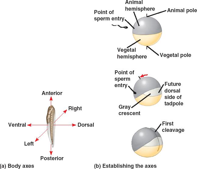
In the frog blastula, the blastocoel is displaced toward the animal pole and is surrounded by a wall several cells thick.
The cell movements that begin gastrulation occur on the dorsal side of the blastula, where the gray crescent was located in
the zygote.
Although still visible as gastrulation begins, the gray crescent is not shown here.
gastrulation-urchin.html: 47_11UrchinGastrulat.jpg
Gastrulation in a sea urchin embryo.
Gastrulation begins with the migration of mesenchyme (mesoderm) cells
from the vegetal pole into the blastocoel.
The vegetal plate invaginates (buckles inward).
Endoderm cells form the archenteron.
Filopodia (made of mesenchyme cells) drag the
archenteron across the blastocoel.
Fusion of the archenteron with the blastocoel wall forms a
digestive tube with a mouth and an anus.
organogenesis-chick.html: 47_15ChickOrganogenesis.jpg
Organogenesis in a chick embryo.
- Early organogenesis.
The archenteron forms when lateral folds pinch the embryo away from the yolk.
The notochord, neural tube, and somites form much as they do in the frog.
- Late organogenesis.
Rudiments of most major organs have already formed in this chick embryo, which is about 56 hours old and about 2–3 mm long.
organogenesis-frog.html: 47_14FrogOrganogenesis_CL.jpg
| Early organogenesis in a frog embryo. |
The dorsal ectoderm folds to form the neural plate.
|
|
The neural plate pinches off to generate the neural tube,
which will become the central nervous system - the brain and the spinal cord.
|
|
The mesoderm forms tissue layers that line the coelom. The somites
will give rise to segmental structures such as vertebrae.
|